Home
Uncategories
Leg Muscles Diagram Posterior : Anatomy Lab Unit 2 Posterior Thigh Muscles Etc Diagram Quizlet / This tutorial teaches the muscles comprising the posterior compartment of the leg.
Leg Muscles Diagram Posterior : Anatomy Lab Unit 2 Posterior Thigh Muscles Etc Diagram Quizlet / This tutorial teaches the muscles comprising the posterior compartment of the leg.
Leg Muscles Diagram Posterior : Anatomy Lab Unit 2 Posterior Thigh Muscles Etc Diagram Quizlet / This tutorial teaches the muscles comprising the posterior compartment of the leg.. Leg muscle anatomy posterior leg muscles diagram photo album 10 / 10 ( 2 votes ) in this image, you will find tensor fascia latae, rectus femoris, vastus lateralis, iliopsoas, pectineus, adductor longus, gracilis, sartorius, vastus medialis, gluteus maximus, adductor magnus, semitendinosus, gracilis. 5 photos of the posterior leg muscles diagram. Leg muscle diagram muscles of the hips and thighs human anatomy and physiology lab. Internal anatomy cross sections of the leg anterior compartment femur nerves & blood vessels posterior compartment 1st metatarsal flexor digitorum longus interosseous artery & medial dorsal. Different types of muscles of arm diagram.
It could be due to soft tissue injury. It contains the plantar flexors: However, many of the leg muscles share functions with other leg muscles. This tutorial is in two parts, the second part is on the muscles of the anterior and lateral compartments of the leg, so please watch that as well! Muscles, arteries, and ne… category:
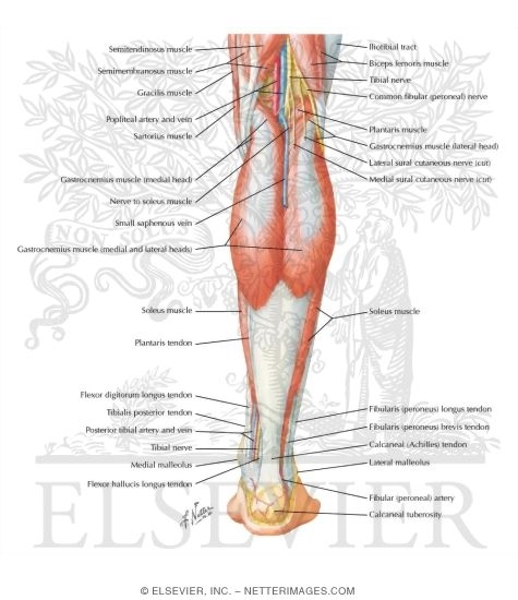
Muscles Of Leg Superficial Dissection Posterior View from netterimages.com Leg muscles can be divided into 3 compartments: The muscle forms the floor of the popliteal fossa the muscle fibers extend through the posterior compartment of the leg and converge to form a solid tendon that passes behind the distal end of the. Have a product modelling and rendering project?. Get a handful labeled leg muscle diagrams to assist your study about human's leg muscle anatomy. Posterior compartment muscles of right lower leg. Anterior compartment, posterior compartment and lateral compartment. It includes the tibialis posterior, the flexor digitorum longus and the flexor. The muscle groups can work independently for specific movements.
Lateral, intermediate and medial cuneiform bone, tuberosity of navicular bone.
Some are small in length, and others are thinner and less bulky than right extensor digitorum longus. Quickly memorize the terms, phrases and much more. Leg muscles functions to perform all the motions and movements of the lower limb like standing, running, dancing etc. Its action causes plantar flexion and inversion of. Lateral, intermediate and medial cuneiform bone, tuberosity of navicular bone. Posted on december 24, 2018december 24, 2018. Leg muscle anatomy posterior leg muscles diagram photo album 10 / 10 ( 2 votes ) in this image, you will find tensor fascia latae, rectus femoris, vastus lateralis, iliopsoas, pectineus, adductor longus, gracilis, sartorius, vastus medialis, gluteus maximus, adductor magnus, semitendinosus, gracilis. Different types of muscles of arm diagram. 3d medical illustration and rendering on leg posterior muscles for our client in australia. Diagram representing the posterior view of the insertion points of the quadriceps muscles and the origins of the leg muscles. Cram.com makes it easy to get the o: The difference comes down to the size of your abdominal muscle bellies. It contains the plantar flexors:
Different types of muscles of arm diagram. Posterior compartment muscles of right lower leg. Create healthcare diagrams like this example called leg muscles in minutes with smartdraw. Muscles of the leg include muscles of the thigh and foot. The sacrum bone is almost always noticeable, no matter what the body type, because it is not covered with muscles or substantial fatty tissue.
Posterior Leg Anatomy Anatomy Drawing Diagram from www.anatomynote.com Its action causes plantar flexion and inversion of. The posterior compartment of the leg contains seven muscles, organized into two layers: Learn vocabulary, terms and more with flashcards, games and other study tools. It could be due to soft tissue injury. Have a product modelling and rendering project?. Leg muscle diagram chapter 13 posterior leg muscles diagram quizlet. The two layers are separated by a band of fascia. Posterior surface fibula, interosseous membrane of leg, surface tibia.
The posterior compartment of the leg is supplied by the tibial nerve.
Leg muscle anatomy posterior leg muscles diagram photo album 10 / 10 ( 2 votes ) in this image, you will find tensor fascia latae, rectus femoris, vastus lateralis, iliopsoas, pectineus, adductor longus, gracilis, sartorius, vastus medialis, gluteus maximus, adductor magnus, semitendinosus, gracilis. The posterior compartment of the leg is supplied by the tibial nerve. Tibialis posterior originates on the proximal 2/3 of tibia and fibula and inserts onto the medial cuneiform and navicular. Leg posterior 3d illustration project. Study flashcards on posterior leg muscles at cram.com. Have a product modelling and rendering project?. Dissection of right lateral cervical region diagram. Anterior compartment, posterior compartment and lateral compartment. 3d medical illustration and rendering on leg posterior muscles for our client in australia. Posted on december 24, 2018december 24, 2018. Leg muscle diagram muscles of the hips and thighs human anatomy and physiology lab. It could be due to soft tissue injury. Posterior muscles in the body.
Posterior view of a left leg, mapping the location of the different muscles that make it up. However, many of the leg muscles share functions with other leg muscles. It contains the plantar flexors: Occurs following excessive exertion of the muscles of anterior compartment of leg. Female hip and leg muscles labeled posterior view, 3d rendering.
Posterior Thigh from www.wesnorman.com Posterior compartment muscles of right lower leg. The posterior compartment of the leg is one of the fascial compartments of the leg and is divided further into deep and superficial compartments. 5 photos of the posterior leg muscles diagram. Posterior surface fibula, interosseous membrane of leg, surface tibia. The difference comes down to the size of your abdominal muscle bellies. This muscle diagram is interactive: Our muscles of the leg quizzes and labeled diagrams are the best way to consolidate your knowledge. Learn vocabulary, terms and more with flashcards, games and other study tools.
Posterior surface fibula, interosseous membrane of leg, surface tibia.
Posterior muscles, such as the hamstrings and gluteus maximus, produce the opposite motion — extension of the thigh at the hip and flexion of the leg at the knee. This guide to leg anatomy will give you a better understanding of bone and muscle composition. Leg muscles anatomy leg anatomy muscle anatomy anatomy study anatomy reference art reference anatomy drawing muscular system your deltoid is made up of three main sets of muscle fibers: Female hip and leg muscles labeled posterior view, 3d rendering. Our muscles of the leg quizzes and labeled diagrams are the best way to consolidate your knowledge. The posterior compartment of the leg contains seven muscles, organized into two layers: The posterior compartment of the leg is supplied by the tibial nerve. 2003 ford escape rear drum brake diagram. Knee muscles, posterior leg muscles anatomy, posterior thigh muscles. Anatomy muscle 3d illustration 3d rendering adductor magnus anatomical arthritis back biceps femoris body buttocks calf muscle diagram female fitness gastrocnemius glutes gluteus maximus gracilis. Study flashcards on posterior leg muscles at cram.com. The muscle groups can work independently for specific movements. It contains the plantar flexors:
The deep muscles that impact leg movement are generally smaller that those that are directly involved in flexing the knee leg muscles diagram. Internal anatomy cross sections of the leg anterior compartment femur nerves & blood vessels posterior compartment 1st metatarsal flexor digitorum longus interosseous artery & medial dorsal.


0 Comments:
Posting Komentar