Home
Uncategories
Anatomy Muscles Pelvis / Anatomical Model Showing The Human Pelvis Muscles Stock Photo Alamy : Then we'll look at the complex sheet of muscles, collectively called the pelvic diaphragm, which form the floor of the pelvic cavity.
Anatomy Muscles Pelvis / Anatomical Model Showing The Human Pelvis Muscles Stock Photo Alamy : Then we'll look at the complex sheet of muscles, collectively called the pelvic diaphragm, which form the floor of the pelvic cavity.
Anatomy Muscles Pelvis / Anatomical Model Showing The Human Pelvis Muscles Stock Photo Alamy : Then we'll look at the complex sheet of muscles, collectively called the pelvic diaphragm, which form the floor of the pelvic cavity.. These muscles all serve as adductors of the thigh, but also serve as important stabilizers of the pelvis and work to maintain balance of the pelvis on the lower limb during gait. Time to solidify your knowledge on the anatomy of the. Attached to the pelvis are muscles of the buttocks, the lower back, and the thighs. These muscles arise from the hip, spine, and proximal femur. These muscles, including the gluteus maximus and the hamstrings, extend the thigh at the hip in support of the body's weight and propulsion.
The many muscles of the hip provide movement, strength, and stability to the hip joint and the bones of the hip and thigh. The muscles of the femoral region of the lower limb are divided into three compartments. Muscles that attach from the pelvis to the trunk and cross the lumbosacral joint muscles that attach from the pelvis to the thigh/leg and cross the hip joint pelvic floor muscles that are located wholly within the pelvis The medial compartment is made up of the adductor magnus, adductor longus, adductor brevis, gracilis and obturator externus. Rectus femoris muscle, one of the quadriceps muscles on the front of your thigh.
Anatomy Model Male Pelvis Ligaments Organs from cdn11.bigcommerce.com Ligaments, tendons, and muscles play an important role in the function of the hip. Each compartment is separated from the others by an intermuscular septum that runs from the fascia lata to the linea aspera of the femur. Piriformis and obturator internus are both hip rotator muscles, which arise within the pelvis, and pass outward through the sciatic foramina. Use the mouse scroll wheel to move the images up and down alternatively use the tiny arrows (>>) on both side of the image to move the images.>>) on both side of the image to move the images. Beginning at the pelvis and running posteriorly along the length of the femur, the majority muscles within the hamstring complex cross both the femoroacetabular and tibiofemoral joints. The iliopsoas muscle consists of the iliac muscle, which comes from the inner surface of the ilium in the pelvis, and the psoas muscle, which originates from the vertebral column. The pelvis's frame is made up of the bones of the pelvis, which connect the axial skeleton to the femurs, and therefore acts in weight bearing of the upper body. Pelvis anatomy muscle thigh muscular system pelvis text hand human png pngwing from w7.pngwing.com this mri pelvis cross sectional anatomy tool is absolutely free to use.
Each compartment is separated from the others by an intermuscular septum that runs from the fascia lata to the linea aspera of the femur.
The levator ani muscles consist of three. Psoas consists of a pair of deep muscles (psoas major and iliacus) located on each side of the pelvis in the abdomen. They form a large sheet of skeletal muscle that is thicker in some areas than in others. Piriformis the piriformis is a triangular muscle 1 on either side on the very front of the posterior wall of true pelvis. Time to solidify your knowledge on the anatomy of the. The function of the pelvic floor is to help assist with child birth, prevent incontinence and support organs within the pelvis. These two muscles join each other and then attach to the lesser trochanter. Attached to the pelvis are muscles of the buttocks, the lower back, and the thighs. These muscles have attachments to the pelvis as follows: The pubococcygeus (pc) muscle is the muscle that runs the show in pelvic floor health. They have several functions, including helping to support the pelvic organs. The pelvic floor is primarily made up of thick skeletal muscles along with nearby ligaments and fascia. These muscles, including the gluteus maximus and the hamstrings, extend the thigh at the hip in support of the body's weight and propulsion.
The semitendinosus, semimembranosus, and biceps femoris muscles comprise the hamstring muscle group. The pubococcygeus (pc) muscle is the muscle that runs the show in pelvic floor health. The muscles of the pelvic floor are collectively referred to as the levator ani and coccygeus muscles. Use the mouse scroll wheel to move the images up and down alternatively use the tiny arrows (>>) on both side of the image to move the images.>>) on both side of the image to move the images. The four groups are the anterior group, the posterior group, adductor group.
5 Facts About The Anatomy Of The Pelvic Cavity from www.visiblebody.com This mri male pelvis axial cross sectional anatomy tool is absolutely free to use. Muscles play an important role in the. The hip joint is one of the most flexible joints in the entire human body. On the posterior side they are the glutei and on the anterior side the hip muscles extending into the thighs. They are also known as the inner hip muscles and deep external rotators. Use the mouse scroll wheel to move the images up and down alternatively use the tiny arrows (>>) on both side of the image to move the images.>>) on both side of the image to move the images. These muscles arise from the hip, spine, and proximal femur. Piriformis the piriformis is a triangular muscle 1 on either side on the very front of the posterior wall of true pelvis.
(1) the obturator internus and the piriformis, which are muscles of the lower extremity, and will be described with these (pages 476 and 477);
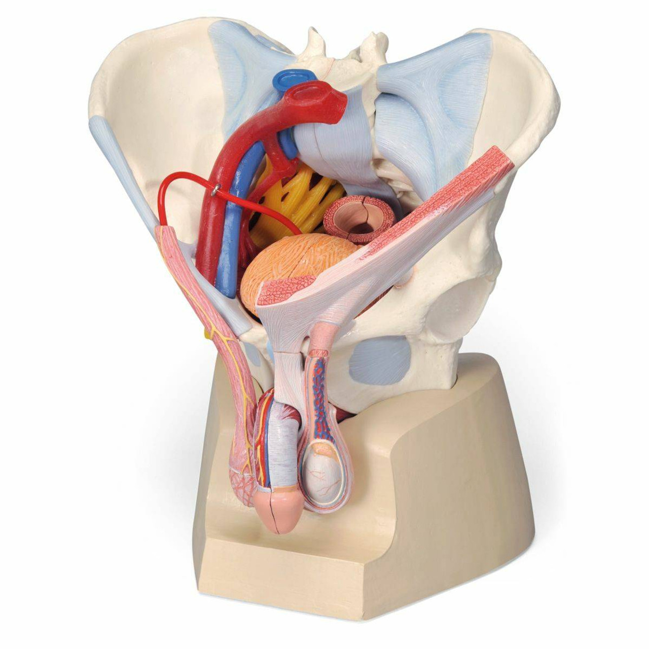
The anterior or extensor, medial or adductor, and posterior or flexor compartments. On the posterior side they are the glutei and on the anterior side the hip muscles extending into the thighs. We'll look at these structures first in a male specimen. (1) the obturator internus and the piriformis, which are muscles of the lower extremity, and will be described with these (pages 476 and 477); The levator ani muscles consist of three. The pelvis's frame is made up of the bones of the pelvis, which connect the axial skeleton to the femurs, and therefore acts in weight bearing of the upper body. The anterior compartment includes pectineus, iliopsoas, psoas minor, iliacus. Then we'll look at the complex sheet of muscles, collectively called the pelvic diaphragm, which form the floor of the pelvic cavity. The floor of the pelvis is formed by the two muscles named levator ani and coccygeus. To support the abdominal and pelvic viscera These two muscles join each other and then attach to the lesser trochanter. The muscles of the pelvis and hip control the vast range of movement of the legs and torso. These muscles originate near the anteroinferior external surface of the bony pelvis and insert at the linea aspera.
These muscles all serve as adductors of the thigh, but also serve as important stabilizers of the pelvis and work to maintain balance of the pelvis on the lower limb during gait. The pelvic floor muscles include; These muscles origin in continuity from the body of the pubis, along a tendinous arch over the obturator internus fascia, and the ischial spine. The iliopsoas muscle consists of the iliac muscle, which comes from the inner surface of the ilium in the pelvis, and the psoas muscle, which originates from the vertebral column. This mri male pelvis axial cross sectional anatomy tool is absolutely free to use.
Normal Anatomy And Physiology Of The Female Pelvis Radiology Key from i0.wp.com To support the abdominal and pelvic viscera It is composed of three separate paired muscles; The anterior compartment includes pectineus, iliopsoas, psoas minor, iliacus. March 23, 2018 anatomy, pelvis and perineum boundaries of pelvic inlet, boundaries of pelvic outlet, boundaries of true pelvis, coccygeus, functions of pelvic diaphragm, iliococcygeus, levator ani muscle, muscles of true pelvis, obtrutor internus muscle, openings in pelvic diaphragm, pelvic diaphragm, pelvic fascia, piriformis muscle, puborectalis, pucococcygeus, question on pelvic diaphragm The levator ani is a broad sheet of muscle. Piriformis the piriformis is a triangular muscle 1 on either side on the very front of the posterior wall of true pelvis. The main function of the pelvic floor muscles are: (1) the obturator internus and the piriformis, which are muscles of the lower extremity, and will be described with these (pages 476 and 477);
These muscles originate near the anteroinferior external surface of the bony pelvis and insert at the linea aspera.
It can be divided into the greater pelvis and the lesser pelvis. The semitendinosus, semimembranosus, and biceps femoris muscles comprise the hamstring muscle group. These muscles arise from the hip, spine, and proximal femur. The iliopsoas muscle consists of the iliac muscle, which comes from the inner surface of the ilium in the pelvis, and the psoas muscle, which originates from the vertebral column. The levator ani is a broad sheet of muscle. We'll look at these structures first in a male specimen. (1) the obturator internus and the piriformis, which are muscles of the lower extremity, and will be described with these (pages 476 and 477); Ligaments, tendons, and muscles play an important role in the function of the hip. The function of the pelvic floor is to help assist with child birth, prevent incontinence and support organs within the pelvis. The many muscles of the hip provide movement, strength, and stability to the hip joint and the bones of the hip and thigh. The floor of the pelvis is formed by the two muscles named levator ani and coccygeus. Arcus tendineus levator ani and the ischial spine The muscles of the femoral region of the lower limb are divided into three compartments.

0 Comments:
Posting Komentar